
Archaeology: The Medium Between the Living and the Dead
By: Claire Gong
Archaeology is often touted as one of the most interdisciplinary subjects. Due to the fragmented and disparate nature of excavated physical evidence, archaeologists must combine different approaches to fit the jigsaw pieces together (Smith, 2019). A prime example of their ingenuity is their incorporation of concepts in medicine into artifact analysis. Although medicine and archaeology are ostensibly dichotomous — the former being concerned with the treatment of disease in the living and the latter aiming to decipher the clues left behind by the dead to reveal the secrets of the past — great breakthroughs have in fact been achieved through the application of medical technology in archaeology. From X-rays and CT scans to PCR testing, medical techniques have transformed the processes of identification, imaging, and analysis of artifacts.
The way archaeology, the study of the dead, harnesses the power of life-saving technology, one could call it a necromancer.
The world first caught a glimpse of the synergy between medicine and archaeology in 1898 when German scientist Walter Konig decided to X-ray a mummified child and cat (Hughes, 2011). The novel X-ray technique enabled non-invasive analysis of body cavities for the first time in history. Less than a decade later, in 1977, the first CT scan of a mummy was conducted by Derek Harwood-Nash in Toronto, Canada (Hughes, 2011). Now, radiological analysis is considered one of the most useful noninvasive techniques (Chhem & Brothwell, 2008). In the case of autopsies, it allows the determination of sex, age, physical characteristics, and pathologies (Lynnerup, 2007). For artifacts, it enables the assessment of morphology and integrity, which generates vital data for artifact preservation and restoration, especially in the case of heavily concreted pieces (Smith, 2019).
There are two types of CT scans: the fan beam and the cone beam CT scan. The traditional fan beam method slices the object into countless sections (Fig. 1.), analogous to slicing a loaf of bread (Anderson & Fregni, 2009). The cone beam, on the other hand, subjects the object to conical radiation (Fig. 2) (Anderson & Fregni, 2009). Cone beam CT scans yield higher spatial resolution, but falls short of fan beam CT scans in terms of the production of clear and anatomically accurate imaging (Lechuga & Weidlich, 2016). Both forms of CT scan generate a wealth of data that is processed by software to produce 3D images that can be viewed from all angles (Anderson & Fregni, 2009).
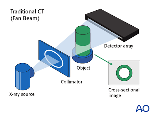
Figure 1. This figure illustrates the mechanism of fan beam CT scans. Image from Schramm et al. 2020.
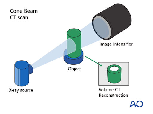
Figure 2. This figure illustrates the mechanism of cone beam CT scans. Image from Schramm et al. 2020.
Genetic testing is another prominent example of the application of medical technology in archaeology. Genetic testing is conducted via polymerase chain reaction (PCR), which amplifies fragments of DNA collected from artifacts or the hard tissues of skeletal remains (Pȁȁbo, 1991). PCR testing only requires small amounts of DNA, making it particularly useful in archaeology, as the autolytic degradation of nucleic acids post-death of organisms leaves only small samples of DNA available (Pȁȁbo, 1991). Since DNA mutations (random changes in genetic base sequence) occur at a relatively constant rate, this “genetic clock” can be used to compare the differences in DNA sequence across various organisms and approximate the time period in which their most recent common ancestor lived (Smith, 2019). PCR testing has enhanced our understanding of our relationship with our ape relatives; for example, we share 98.8% genetic homologies with chimpanzees (Smith, 2019). It has also elucidated the mystery of human origins. Genetic testing revealed that genetic diversity is highest in Africa and decreases as we move away, which corroborates the out-of-Africa model (Cann et al., 1987) and indicates a series of population bottlenecks (extreme reductions in population size, for example, due to natural disasters). These bottlenecks potentially reduced variation within the gene pool, explaining the surprisingly low amount of genetic diversity within the human population (Foley, 1994).
The utility of medical technologies, such as X-rays, CT scanning, and PCR testing within the field of archaeology demonstrates its brilliant chemistry with medicine. It impressively exemplifies how the whole is more than the sum of its parts, showing the magic that happens when two dichotomous disciplines collide. As archaeologists continue to engage in interdisciplinary exploration, we can only imagine what fascinating discoveries are in store for us.
References
Anderson, G., & Fregni, G. (2009). Technology as a tool for archaeological research and artifact conservation. Objects Specialty Group Postprints, 16, 95-109. http://29aqcgc1xnh17fykn459grmc-wpengine.netdna-ssl.com/osg-postprints/wp-content/uploads/sites/8/2015/02/osg016-08.pdf
Cann, R. L., Stoneking, M., & Wilson, A. C. (1987). Mitochondrial DNA and human evolution. Nature, 325(6099), 31-36. 10.1038/325031a0
Foley, R. A. (1994). Speciation, extinction and climatic change in hominid evolution. Journal of Human Evolution, 26(4), 275-289. https://doi.org/10.1006/jhev.1994.1017
Hughes, S. (2011). CT Scanning in Archaeology. ()10.5772/22741
Lechuga, L., & Weidlich, G. A. (2016). Cone Beam CT vs. Fan Beam CT: A Comparison of Image Quality and Dose Delivered Between Two Differing CT Imaging Modalities. Cureus, 8(9), e778. 10.7759/cureus.778
Lynnerup, N. (2007). Mummies. American Journal of Physical Anthropology, 134, 162-190. https://doi.org/10.1002/ajpa.20728
O'Connor, S. (2008). Paleoradiology: imaging mummies and fossils. Rethy K. Chhem & Don R. Brothwell. Springer-Verlag: Berlin and Heidelberg, 2008. 163 pp. ISBN 10 3540488324. International Journal of Osteoarchaeology, 18(5), 546-548. https://doi.org/10.1002/oa.1016
Pääbo, S. (1991). Amplifying DNA from archeological remains: a meeting report. PCR Methods and Applications, 1(2), 107-110.
Schramm, A., Metzger, M., Gellrich, N. & Strong, B. (2020). Cone beam vs fan beam CT. Surgery Reference. https://surgeryreference.aofoundation.org/cmf/further-reading/cas-cone-beam-vs-fan-beam-ct
Smith, C. (2019). Encyclopedia of global archaeology. https://doi.org/10.1007/978-3-319-51726-1
Zesch, S., Panzer, S., Rosendahl, W., Nance,John W.,,Jr, Schönberg, S.,O., & Henzler, T. (2016). From first to latest imaging technology: Revisiting the first mummy investigated with X-ray in 1896 by using dual-source computed tomography. European Journal of Radiology Open, 3, 172-181. 10.1016/j.ejro.2016.07.002
Images
Schramm, A., Metzger, M., Gellrich, N. & Strong, B. (2020). Traditional CT (Fan Beam). Surgery Reference. https://surgeryreference.aofoundation.org/cmf/further-reading/cas-cone-beam-vs-fan-beam-ct
Schramm, A., Metzger, M., Gellrich, N. & Strong, B. (2020). Cone Beam CT Scan. Surgery Reference. https://surgeryreference.aofoundation.org/cmf/further-reading/cas-cone-beam-vs-fan-beam-ct